
What Is Calcific Tendinitis of the Shoulder?
Calcific tendinitis of the shoulder is a common cause of sudden shoulder pain, especially when calcium deposits form inside the rotator cuff tendons.
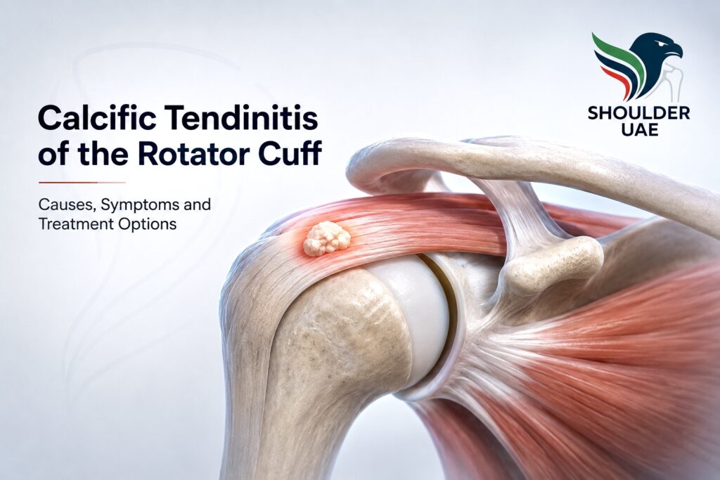
The rotator cuff is a group of four tendons that help lift, rotate and stabilize the shoulder. In calcific tendinitis, calcium crystals — usually hydroxyapatite — accumulate within the tendon substance. The supraspinatus tendon is the most commonly affected, although other tendons such as the infraspinatus or subscapularis may also be involved.
For some patients, the calcium deposit is found incidentally on an X-ray and causes no symptoms. For others, it can produce sudden, intense shoulder pain, night pain, stiffness and difficulty lifting the arm.
The good news is that many patients improve without surgery. However, accurate diagnosis is important because calcific tendinitis can mimic other shoulder problems, including rotator cuff tears, frozen shoulder, bursitis and impingement syndrome.
What causes calcific tendinitis?
The exact cause is not always clear. Calcific tendinitis is not simply “too much calcium in the diet” and it is not usually related to drinking milk or taking calcium supplements.
Current understanding suggests that it is an active biological process within the tendon. Tendon cells may temporarily change their behavior and allow calcium crystals to form inside the tendon. This can be influenced by several factors:
- Tendon overload or repetitive shoulder use
- Reduced local blood supply within part of the tendon
- Degenerative or metabolic tendon changes
- Mechanical compression in the subacromial space
- Hormonal or endocrine factors in some patients
- A natural cycle of calcium formation and later resorption
It is most often seen in adults between 30 and 60 years old and is slightly more common in women. It can affect one or both shoulders.
The phases of calcific tendinitis

Understanding the phase of calcific tendinitis helps guide treatment and explains why shoulder pain can vary over time.
1. Pre-calcific phase
In this early phase, tendon cells begin to change, creating the environment where calcium can later be deposited. Patients may have no symptoms or only mild discomfort.
2. Formative phase
Calcium begins to form inside the tendon. The deposit is often dense and well-defined on X-ray. Pain may be mild, moderate or absent. Some patients notice pain when lifting the arm, reaching overhead or lying on the affected side.
3. Resting phase
The calcium deposit remains within the tendon but may not be very inflamed. Symptoms can fluctuate. Some patients live with the deposit for months or even years without major pain.
4. Resorptive phase
This is often the most painful phase. The body starts to break down and absorb the calcium deposit. The deposit becomes softer and more “toothpaste-like”. This phase can trigger a strong inflammatory reaction in the tendon and surrounding bursa.
Patients may experience sudden severe pain, marked night pain, inability to lift the arm and stiffness. This is the phase that often brings patients urgently to a shoulder specialist.
5. Post-calcific or healing phase
After the calcium has been resorbed or removed, the tendon gradually heals. Pain improves and shoulder movement returns. Rehabilitation may be needed to recover full motion, strength and confidence.
Common symptoms

Calcific tendinitis can present in different ways. Symptoms may be gradual or sudden.
Common symptoms include:
- Pain on the side or front of the shoulder
- Pain radiating into the upper arm
- Severe night pain
- Difficulty sleeping on the affected shoulder
- Pain when lifting the arm overhead
- Pain with dressing, reaching behind the back or fastening a seatbelt
- Loss of range of motion
- A feeling of stiffness or “frozen shoulder”
- Weakness due to pain inhibition
In acute cases, the pain can be very intense and may occur without trauma. Some patients are worried because they suddenly cannot move the shoulder, even though there has been no injury.
Is calcific tendinitis the same as a rotator cuff tear?

No. Calcific tendinitis and rotator cuff tear are different conditions, but they can produce similar symptoms.
In calcific tendinitis, the main problem is a calcium deposit inside the tendon. In a rotator cuff tear, the tendon fibers are partially or completely torn.
However, both conditions can coexist. A patient with a calcium deposit may also have tendon degeneration, partial tearing or a full-thickness rotator cuff tear. This is why accurate imaging is important, especially when there is weakness, persistent pain, previous trauma or symptoms that do not improve as expected.
Before deciding treatment, the shoulder specialist should assess:
- The size and location of the calcium deposit
- Whether the deposit is hard or soft
- The degree of bursitis or inflammation
- Tendon quality
- Whether there is a partial or complete rotator cuff tear
- Shoulder stiffness
- The patient’s activity level and expectations
How is calcific tendinitis diagnosed?

Diagnosis starts with a careful clinical examination. The specialist will assess pain location, range of motion, strength, impingement signs, stiffness and possible associated conditions.
X-ray
A shoulder X-ray is often the first imaging test. It can show the calcium deposit clearly and help assess its size, shape and location.
Ultrasound
High-resolution ultrasound is very useful because it allows dynamic assessment of the rotator cuff. It can show the calcium deposit, bursitis, tendon inflammation and many rotator cuff tears. It also allows image-guided treatment, such as injection or barbotage, when indicated.
MRI
MRI may be recommended when symptoms are persistent, when there is significant weakness, when a rotator cuff tear must be excluded, or when surgery is being considered. MRI can show tendon tears, bursitis, bone edema, inflammation and other shoulder pathology.
Treatment options for calcific tendinitis

Treatment depends on the severity of symptoms, the phase of the calcification, the size and type of deposit, and whether there is an associated rotator cuff tear.
Most patients start with non-surgical treatment.
1. Activity modification and pain control
During painful episodes, avoiding aggravating activities is important. This does not mean complete immobilization for a long time. The aim is to reduce pain while maintaining gentle movement to avoid stiffness.
Treatment may include:
- Avoiding repetitive overhead activity
- Ice or heat depending on comfort
- Short-term anti-inflammatory medication if medically appropriate
- Pain medication when needed
- Gentle range-of-motion exercises
Anti-inflammatory medication should be used carefully in patients with stomach problems, kidney disease, blood thinners, cardiovascular risk or other medical conditions.
2. Physiotherapy
Physiotherapy is useful once the severe pain settles. In the acute inflammatory phase, aggressive strengthening may worsen symptoms. The first goal is usually to reduce pain and restore movement.
A structured rehabilitation program may include:
- Gentle mobility exercises
- Posterior capsule stretching if needed
- Scapular control
- Rotator cuff strengthening
- Postural correction
- Progressive return to sport or gym activity
Physiotherapy is particularly important when calcific tendinitis causes secondary stiffness or altered shoulder mechanics.
3. Ultrasound-guided subacromial injection
If pain is severe or sleep is affected, an ultrasound-guided injection into the subacromial bursa may help reduce inflammation and pain.
The injection does not remove the calcium deposit, but it can calm the inflammatory reaction around it. Ultrasound guidance improves accuracy and helps place the medication exactly where it is needed.
This option can be useful in acute painful episodes, especially when there is associated bursitis.
4. Shockwave therapy
Extracorporeal shockwave therapy uses focused mechanical energy applied from outside the body. It may help reduce pain, stimulate local healing and promote calcium deposit fragmentation or resorption.
Shockwave therapy is non-surgical and can be considered for persistent calcific tendinitis, especially when symptoms continue despite initial treatment.
It is not suitable for every patient and results can vary depending on the type, size and chronicity of the deposit.
5. Ultrasound-guided barbotage or lavage
Barbotage is a minimally invasive procedure performed under ultrasound guidance. A needle is placed precisely into the calcium deposit. Saline is then used to break down and wash out the calcium material.
This technique is usually more effective when the deposit is soft or in the resorptive phase. Very hard, dense calcifications may be more difficult to aspirate.
Barbotage may be combined with a subacromial injection to reduce inflammation after the procedure.
Potential benefits include:
- Direct treatment of the calcium deposit
- Minimally invasive approach
- Real-time ultrasound guidance
- Faster pain relief in selected patients
- Avoidance of surgery in many cases
However, not every calcification requires barbotage. Treatment should be individualized after clinical examination and imaging.
6. Arthroscopic surgery

Surgery is not the first treatment for most patients with calcific tendinitis. It may be considered when pain persists despite appropriate non-surgical treatment, when the deposit is large and mechanically symptomatic, or when there is an associated rotator cuff tear that needs repair.
Shoulder arthroscopy is a minimally invasive surgical technique performed through small incisions using a camera and specialized instruments.
During arthroscopy, the surgeon can:
- Inspect the entire shoulder joint
- Remove the calcium deposit
- Treat inflamed bursal tissue
- Assess the rotator cuff tendon from both sides
- Repair the rotator cuff if there is a significant tear
- Treat associated pathology if present
If removing the calcium leaves a tendon defect, the tendon may need to be repaired. This is one of the key reasons why calcific tendinitis should be managed by a shoulder specialist familiar with both ultrasound-guided procedures and arthroscopic rotator cuff surgery.
When should you see a shoulder specialist?

You should consider seeing a shoulder specialist if you have:
- Severe shoulder pain without trauma
- Pain that wakes you at night
- Inability to lift the arm
- Symptoms lasting more than a few weeks
- Recurrent episodes of shoulder pain
- Weakness suggesting a rotator cuff tear
- Stiffness that is getting worse
- Previous treatment without improvement
- A known calcium deposit on X-ray or ultrasound
Early assessment helps identify the correct diagnosis and avoid unnecessary delays.
Can calcific tendinitis come back?
In many patients, once the calcium deposit resolves, symptoms improve significantly. Recurrence can happen, but it is not inevitable.
Persistent pain after the calcium has disappeared may be due to:
- Residual bursitis
- Shoulder stiffness
- Rotator cuff tendinopathy
- Partial tendon tear
- Frozen shoulder
- AC joint arthritis
- Neck-related pain
This is why follow-up is important if symptoms do not match the imaging findings.
Recovery and return to activity

Recovery depends on the phase and severity of the condition.
Mild cases may improve within a few weeks. More painful or chronic cases may take several months. After barbotage, patients often need a short period of relative rest followed by physiotherapy. After arthroscopy, recovery depends on whether the tendon required repair.
As a general principle:
- Pain control comes first
- Range of motion should be restored early but gently
- Strengthening should be progressive
- Return to gym, swimming, tennis, padel or overhead sport should be guided by symptoms, strength and tendon status
Trying to “push through” severe pain is usually not helpful and may prolong inflammation or stiffness.
Expert shoulder care in Abu Dhabi
Calcific tendinitis can be extremely painful, but most patients improve with the correct diagnosis and a structured treatment plan.
At Shoulder UAE, Dr. Pablo Menéndez Martínez provides specialist assessment for rotator cuff and tendon disorders, including calcific tendinitis, bursitis, impingement and rotator cuff tears. Treatment options include advanced imaging review, ultrasound-guided procedures, barbotage, injections, rehabilitation planning and minimally invasive shoulder arthroscopy when needed.
The goal is always to reduce pain, restore function and help each patient return safely to daily life, work and sport.
Book an appointment with Dr Pablo Menendez Martinez
Frequently Asked Questions
Is calcific tendinitis dangerous?
No, it is not dangerous in the sense of being cancerous or life-threatening. However, it can be extremely painful and disabling. Proper diagnosis is important to exclude associated problems such as rotator cuff tears or frozen shoulder.
Does calcific tendinitis always need surgery?
No. Most patients improve without surgery. Treatment may include medication, physiotherapy, ultrasound-guided injection, shockwave therapy or barbotage. Surgery is reserved for selected cases that do not improve or have associated tendon damage.
Can the calcium deposit disappear by itself?
Yes. Calcific tendinitis can follow a natural cycle in which the body eventually resorbs the calcium. The resorptive phase can be very painful but may also indicate that the deposit is breaking down.
Is barbotage painful?
Barbotage is usually performed with local anesthetic and ultrasound guidance. Patients may feel pressure or discomfort during the procedure, and the shoulder can be sore for a few days afterwards.
How do I know if I have a rotator cuff tear as well?
Clinical examination and imaging help answer this. Ultrasound and MRI can assess tendon integrity. A rotator cuff tear is more likely if there is true weakness, history of trauma, older age, or persistent symptoms despite treatment.
Can I exercise with calcific tendinitis?
Gentle movement is usually encouraged, but painful overhead lifting, heavy gym exercises or repetitive loading may worsen symptoms during an acute flare. A specialist or physiotherapist can guide a safe exercise plan.
What is the best treatment for calcific tendinitis?
There is no single best treatment for every patient. The best option depends on pain severity, the phase and type of calcification, tendon condition, shoulder stiffness and previous treatments. A personalized plan gives the best chance of recovery.
Further Reading & Medical References
This article is intended for patient education and is based on specialist clinical experience, current shoulder literature and internationally recognised medical resources.
For further reading, you may find the following external resources helpful:
- American Academy of Orthopaedic Surgeons. Calcific Tendinitis of the Shoulder.
- NCBI Bookshelf / StatPearls. Ultrasound-Guided Barbotage.
- The BMJ. Ultrasound guided lavage with corticosteroid injection versus sham lavage for calcific tendinopathy of the shoulder.
- National Library of Medicine / PubMed Central. Diagnosis and treatment of calcific tendinitis of the shoulder.